Diagram Of Shoulder Muscles And Tendons : However, their origin is found in the osseous structures and they are not to be included with the rotator cuff muscles.
Diagram Of Shoulder Muscles And Tendons : However, their origin is found in the osseous structures and they are not to be included with the rotator cuff muscles.. The shoulder joint is formed where the humerus (upper arm bone) fits into the scapula. The rotator cuff is a group of four muscles and tendons that surround the glenohumeral joint. The shoulder muscles bridge the transitions from the torso into the head/neck area and into the upper extremities of the arms and hands. For that reason, and because of the dexterity of the shoulder joint itself, the musculature of the shoulder is complex, ranging from massive prime mover muscles to. The function of this entire muscular apparatus is to produce.
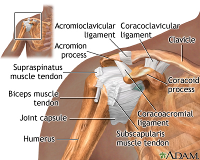
Tendons are extensions of muscles that attach muscles to bone. The shoulder is not a single joint, but a complex arrangement of bones, ligaments, muscles, and tendons that is better called the shoulder girdle. An mri of the shoulder of a healthy subject was performed in the 3 planes of space (coronal, axial, sagittal) commonly used in osteoarticular imaging, with two weightings to explore the musculoskeletal pathology of the shoulder: Specifically, the four rotator cuff muscles include the following Following inferior dislocation of shoulder joint, the rounded contour of shoulder is lost and there is weakness of abduction of armbecause the axillary nerve is likely to be injured in the inferior.
Shoulder Problem Wikipedia from upload.wikimedia.org Muscles move the bones by pulling on the tendons. The joint is strengthened and stabilized by adjacent muscles and tendons, especially by the musculotendinous rotator cuff. Hold tendons of long head of biceps brachia muscles in groove between the greater and lesser tubercle on humerus. Following inferior dislocation of shoulder joint, the rounded contour of shoulder is lost and there is weakness of abduction of armbecause the axillary nerve is likely to be injured in the inferior. Each of these muscles is a discrete organ constructed of skeletal muscle tissue, blood vessels, tendons, and nerves. Joanna and her team specialise in providing custom made illustrations to suit all projects. The human shoulder is made up of three bones: An example of shoulder flexion can be seen when reaching forward to grasp an object.
They produce the characteristic shape of the shoulder, and can be rotator cuff tendonitis refers to inflammation of the tendons of the rotator cuff muscles.
An mri of the shoulder of a healthy subject was performed in the 3 planes of space (coronal, axial, sagittal) commonly used in osteoarticular imaging, with two weightings to explore the musculoskeletal pathology of the shoulder: The rotator cuff is a group of four muscles and tendons that surround the glenohumeral joint. The rotator cuff muscles are a collection of muscles and tendons that surround the shoulder, giving it support and allowing its wide range of motion. This usually occurs secondary to repetitive use of the shoulder. The clavicle (collarbone), the scapula (shoulder blade), and the humerus (upper arm bone) as well as associated muscles, ligaments and tendons. Related posts of shoulder muscles and tendons diagram back muscles anatomy. The muscles of the shoulder are associated with movements of the upper limb. Muscles of the shoulder work in team to produce highly coordinated motion. The large deltoid muscle is the outer layer of shoulder muscle. The function of this entire muscular apparatus is to produce. The muscles of the rotator cuff include the suprasinatus, infraspinatus, teres minor, and subscapularis. Learn vocabulary, terms and more with flashcards, games and other study tools. Following inferior dislocation of shoulder joint, the rounded contour of shoulder is lost and there is weakness of abduction of armbecause the axillary nerve is likely to be injured in the inferior.
Starting point the muscles are the supraspinatus this is a flat triangular muscle that fills the entire infraspinatus fossa. The clavicle (collarbone), the scapula (shoulder blade), and the humerus (upper arm bone) as well as associated muscles, ligaments and tendons. The muscles of the rotator cuff include the suprasinatus, infraspinatus, teres minor, and subscapularis. Tendons are much like ligaments, except that tendons attach muscles to bones. Each of these muscles is a discrete organ constructed of skeletal muscle tissue, blood vessels, tendons, and nerves.
Shoulder Pain Uf Health University Of Florida Health from m.ufhealth.org The shoulder is one of the largest and most complex joints in the body. It relies on ligaments and muscle tendons to provide reinforcement. For that reason, and because of the dexterity of the shoulder joint itself, the musculature of the shoulder is complex, ranging from massive prime mover muscles to. Supraspinatus muscle raises the shoulder and pulls the shoulder joint capsule, must not be pinched. An mri of the shoulder of a healthy subject was performed in the 3 planes of space (coronal, axial, sagittal) commonly used in osteoarticular imaging, with two weightings to explore the musculoskeletal pathology of the shoulder: Ready to test your knowledge on those muscles? Specifically, the four rotator cuff muscles include the following From simple diagrams to complex anatomy, and is readily able.
The muscles of the rotator cuff include the suprasinatus, infraspinatus, teres minor, and subscapularis.
Tendons are extensions of muscles that attach muscles to bone. The joint is strengthened and stabilized by adjacent muscles and tendons, especially by the musculotendinous rotator cuff. The function of this entire muscular apparatus is to produce. Ready to test your knowledge on those muscles? It relies on ligaments and muscle tendons to provide reinforcement. Starting point the muscles are the supraspinatus this is a flat triangular muscle that fills the entire infraspinatus fossa. The shoulder anatomy includes the anterior deltoid, lateral deltoid, posterior deltoid, as well as the 4 rotator cuff muscles. Shoulder joint allows lifting, pushing and pulling by upper extremity. Shoulder flexion is movement of the shoulder in a forward motion. Each of these muscles is a discrete organ constructed of skeletal muscle tissue, blood vessels, tendons, and nerves. Weakness of any muscle change normal kinematic chain of the joint. The large deltoid muscle is the outer layer of shoulder muscle. The shoulder muscles include skeletal muscles that are attached to the head of the humerus which performs various direct and indirect functions of the shoulder joints.
The capsule is strengthened by the tendons and ligaments surrounding and blending with it. Shoulder joint allows lifting, pushing and pulling by upper extremity. Each of these muscles is a discrete organ constructed of skeletal muscle tissue, blood vessels, tendons, and nerves. The muscles of the rotator cuff include the suprasinatus, infraspinatus, teres minor, and subscapularis. The large deltoid muscle is the outer layer of shoulder muscle.
Shoulder Problem Wikipedia from upload.wikimedia.org Muscles move the bones by pulling on the tendons. The collection of muscles and tendons in the shoulder is known as the rotator cuff. The human shoulder is made up of three bones: Muscles of the shoulder work in team to produce highly coordinated motion. The muscles of the rotator cuff include the suprasinatus, infraspinatus, teres minor, and subscapularis. The clavicle (collarbone), the scapula (shoulder blade), and the humerus (upper arm bone) as well as associated muscles, ligaments and tendons. An mri of the shoulder of a healthy subject was performed in the 3 planes of space (coronal, axial, sagittal) commonly used in osteoarticular imaging, with two weightings to explore the musculoskeletal pathology of the shoulder: The shoulder muscles include skeletal muscles that are attached to the head of the humerus which performs various direct and indirect functions of the shoulder joints.
Because shoulders have such range of movement and are used often, they are commonly injured.
Muscles move the bones by pulling on the tendons. The muscles of the rotator cuff include the suprasinatus, infraspinatus, teres minor, and subscapularis. Webmd's shoulder anatomy page provides an image of the parts of the shoulder and describes its function, shoulder problems, and more. From simple diagrams to complex anatomy, and is readily able. An example of shoulder flexion can be seen when reaching forward to grasp an object. Start studying shoulder ligaments and tendons. The muscles of the shoulder dynamically function in performing a wide range of motion, specifically the rotator cuff muscles which function to move the the rotator cuff (rc) is an anatomic coalescence of the muscle bellies and tendons of the supraspinatus (ss), infraspinatus (is), teres minor (tm). The collection of muscles and tendons in the shoulder is known as the rotator cuff. The rotator cuff tendons are a group of four tendons that connect the deepest layer of muscles to the humerus. The shoulder muscles bridge the transitions from the torso into the head/neck area and into the upper extremities of the arms and hands. The shoulder is one of the largest and most complex joints in the body. The shoulder muscles include skeletal muscles that are attached to the head of the humerus which performs various direct and indirect functions of the shoulder joints. Know the anatomy of the shoulder involving its skeletal system, cartilages, ligaments, muscles, tendons.


0 Comments